
Most patients have seen facelift before and after photographs. Far fewer have seen what actually happens during the operation. In this surgical video, I take you inside the operating room to show how I perform a deep plane facelift, from the initial markings and anesthesia to the deeper tissue dissection, skin closure and neck contouring.
I want patients to understand that a facelift is not simply a matter of removing loose skin and pulling everything tighter. The important work involves understanding the anatomy beneath the skin and repositioning the tissues that have changed with aging.
This seven-minute video follows an actual operation. It is intended to give you a better understanding of the procedure rather than demonstrate every technical detail involved in a facelift.
Viewer discretion: The following video contains actual surgical footage, including incisions, tissue dissection and suturing.
INSIDE THE OPERATING ROOM
Watch Dr. Athré Perform Deep Plane Facelift Surgery
In this narrated surgical demonstration, I explain the markings, anesthesia, facial dissection, SMAS repositioning, incision closure and neck tightening involved in the operation.
UNDERSTANDING THE PROCEDURE
What Happens During Deep Plane Facelift Surgery?
The operation shown in this video has five major stages. Each serves a different purpose, but they all contribute to the final result.
First, I prepare and anesthetize the tissues. I then create the incisions and elevate the skin to expose the deeper facial anatomy. The SMAS and associated muscular tissues are dissected and repositioned before the excess skin is removed. Finally, I complete the neck work and close the incisions.
Understanding the difference between these stages is important because the deeper tissue work is what separates modern structural facelift techniques from simply tightening the skin.
CHAPTER 01
Preparation, Surgical Markings and Anesthesia
Every facelift begins with planning. Before making an incision, I mark the areas where I intend to work. These markings help establish the incision locations and give me an anatomical roadmap during surgery.
The position of an incision matters. I consider the natural contours around the ears, the hairline and the neck so that the surgical approach provides the access I need while allowing the incisions to heal in appropriate locations.
In the video, the patient has already received sedation and major nerve blocks before I begin injecting local anesthetic throughout the face and neck.
The local anesthetic provides additional numbness within the tissues. It also contains epinephrine, which constricts small blood vessels and helps limit bleeding during the initial dissection.
You can see the patient making facial expressions while I work. She appears comfortable during this portion of the procedure. Anesthetic plans are individualized, and this particular approach is not necessarily appropriate for every patient.
CHAPTER 02
Making the Incisions and Elevating the Skin
I begin the surgical dissection with a small incision beneath the chin. This is called a submental incision, and it provides access to the central neck.
Using facelift scissors, I separate the skin from the underlying tissues. The initial dissection takes place in the subcutaneous plane, which simply means beneath the skin.
In this particular operation, I also perform a small amount of liposuction to improve tissue contouring and visualization.
Next, I make the planned incisions around each ear using a No. 15 scalpel blade. The skin is carefully elevated to expose the deeper structures that will be repositioned later in the operation.
Behind the ear, this dissection connects with the area already prepared in the neck. This allows me to work across the lower face and neck while maintaining a coordinated surgical plan.
The first part of the dissection is subcutaneous, but that does not mean the entire operation is a skin-only facelift. It is the preparation that allows me to access the deeper tissues.
Small bleeding vessels are controlled as the operation progresses. In the video, I use bipolar electrocautery before moving to the deeper anatomical layers.
CHAPTER 03
Dissecting and Repositioning the SMAS Layer
This is the portion of the operation I particularly want patients to understand.
After the skin has been elevated and bleeding points have been addressed, I identify and mark important structures on the SMAS, or superficial musculoaponeurotic system.
The SMAS is a fibrous and muscular layer beneath the skin. It is an important part of facial support and a central structure in modern facelift surgery.
I inject saline beneath this layer to help separate tissue planes. This is called hydrodissection. The saline can facilitate elevation of the SMAS from the tissues beneath it.
The subsequent dissection is careful and meticulous. Much of the separation is accomplished by gently spreading the tissues rather than applying excessive force.
With a deep plane facelift, the purpose of working beneath the SMAS is to create greater mobility in selected deeper facial tissues. Depending on the approach, this can include releasing the facial retaining ligaments that restrict movement.
The surgical video is a condensed overview, so it does not identify every individual ligament or anatomical structure encountered during the operation.
In the lateral neck, the dissection continues beneath the platysma muscle. Once the tissues are adequately mobilized, I reposition them in a posterior and superior direction, meaning backward and upward.
I trim excess muscle tissue where necessary to avoid bunching and secure the repositioned tissues with fine sutures.
The important point is that a well-executed dissection allows the tissues to move into their new position without requiring excessive tension.
I am not trying to pull the skin tighter to create the result. I am repositioning the deeper tissues so the skin can follow naturally.
Near the end of this stage, you can see the excess skin remaining after the deeper tissues have been lifted. That is an especially useful moment because it demonstrates why skin removal comes after the structural repositioning.
CHAPTER 04
Removing Excess Skin and Closing the Incisions
Once the deeper tissues have been repositioned, the excess skin becomes apparent. I can now assess how much skin needs to be removed without relying on it to maintain the lift.
In this video, I apply Tisseel, a fibrin tissue sealant, to the surgical bed. It is used as an additional measure in managing bleeding and tissue adherence. It does not eliminate the possibility of postoperative bleeding or hematoma.
One of the most important details in this portion of the operation is the positioning of the earlobe.
I refer to this step as the delivery of the ear lobule.
The earlobe needs to sit naturally at the junction of the face and neck. Excessive tension around this area can create an unnatural, pulled appearance. I pay particular attention to the way the skin is positioned and closed around the ear.
After carefully marking the excess skin, I remove it and close the incisions using meticulous suturing techniques.
The objective is to have the deeper tissues supporting the lift while the skin is redraped and closed with minimal tension. Careful closure helps protect the natural appearance of the ear, although all surgical incisions leave scars that continue to mature during healing.
CHAPTER 05
Completing the Neck Lift
In the final portion of the video, I return to the neck.
I perform a midline platysmaplasty, which involves tightening and repositioning the platysma muscle beneath the chin. This helps improve the relationship between the underside of the chin and the neck.
I then address the excess skin along the posterior and lateral neck.
The face and neck are closely connected anatomically. For many patients, improving the jawline means evaluating more than the tissues immediately around the cheeks and jowls.
Skin laxity, platysma position, fat distribution and deeper neck anatomy can all contribute to the appearance of the neck. This is why the exact combination of procedures varies between patients.
Some patients need more extensive work beneath the platysma. Others may benefit from a more limited approach. My neck lift procedure page explains the anatomical issues involved in improving neck contour.
As I explain at the conclusion of the video, this is an overview of the operation. It is not intended to illustrate every possible facelift or neck lift technique.
SURGICAL RESULTS
How the Surgical Work Translates Into Results
During the operation, you are watching the tissue layers being separated and repositioned. In before and after photographs, you can begin to see how that surgical work translates into changes in the jawline, lower face and neck.
These photographs are from my patient gallery. They are separate cases from the patient shown in the surgical video, and several involve additional facial rejuvenation procedures.
BEFORE & AFTER
Deep Plane Facelift Patient Results
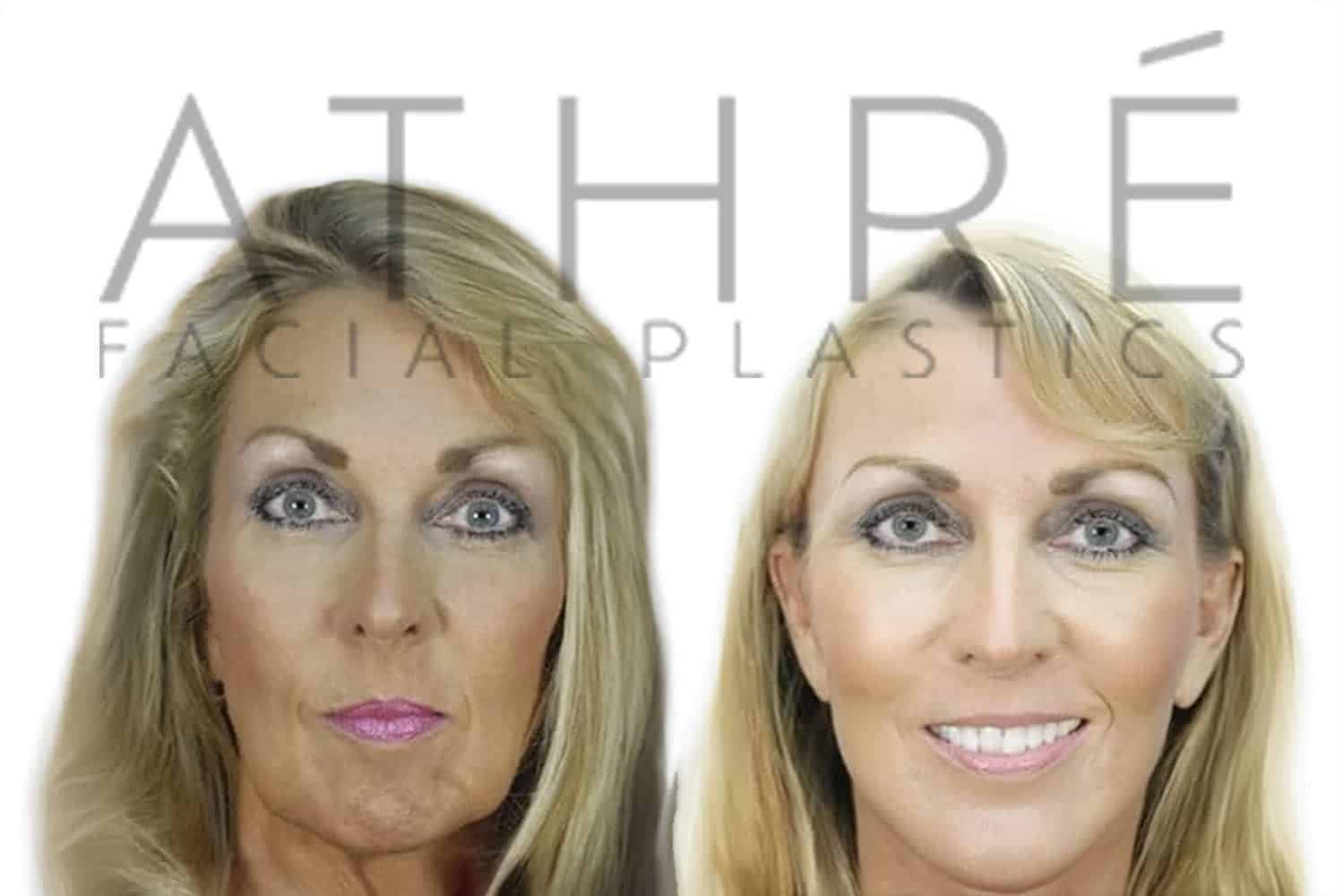


Results vary based on anatomy, the extent of surgery, healing and whether additional procedures were performed. These photographs show real patient outcomes, not a guarantee of what another patient will experience.
DR. ATHRÉ’S PERSPECTIVE
What I Want Patients to Understand About Deep Plane Facelift Surgery
The most important part of a facelift is not how much skin I remove. It is how I mobilize and reposition the tissues that have descended over time.
When the deeper tissues are properly addressed, I can redrape the skin without depending on excessive skin tension to maintain the result. That allows me to focus on the position of the ears, the appearance of the incisions and the overall relationship between the face and neck.
I also want patients to understand that the operation is individualized. Not every patient needs the same degree of dissection, neck work or additional facial rejuvenation procedures.
The seven-minute video demonstrates one surgical approach, but the actual treatment plan begins with examining your anatomy and understanding what you want to change.
The operation is only part of the process. Swelling, bruising, temporary numbness and incision healing continue after surgery. Understanding the postoperative healing process is just as important as understanding what happens in the operating room.
CONTINUE LEARNING WITH DR. ATHRÉ
More About Deep Plane Facelift Surgery
You’ve seen how I perform the operation. These additional short videos explore the decisions behind my technique, the deeper anatomy of the face, incision placement, anesthesia, recovery and longer-term patient results.
FREQUENTLY ASKED
Deep Plane Facelift Surgery FAQs
These are some of the questions patients ask about the operation shown in the video.
What makes this a deep plane facelift?
The operation includes deeper dissection and repositioning involving the SMAS and associated tissues, rather than relying on skin tightening alone. Research describing deep plane anatomy explains the relationships between these layers, retaining ligaments and facial nerves.
Can a deep plane facelift be performed under local anesthesia?
Yes, in appropriately selected patients. The patient in this video received sedation and nerve blocks before local anesthetic infiltration. A review of facelift procedures under local anesthesia discusses patient selection, monitoring and anesthetic techniques.
Why is there an incision beneath the chin?
The submental incision provides access to the central neck, where I perform a midline platysmaplasty in this operation. A review of neck lift anatomy and techniques explains the role of the platysma and deeper structures in neck contouring.
Why is tension-free closure important around the ears?
Excessive skin tension can distort the earlobe or contribute to an unnatural appearance. Published research on earlobe deformity after facelift surgery discusses techniques intended to reduce this problem.
Does Tisseel prevent bleeding after surgery?
No. It is an additional measure, not a guarantee. A meta-analysis of tissue sealants in facelift surgery reported reduced drainage and a possible reduction in hematoma, but careful bleeding control remains essential.
Is a deep plane facelift better than a traditional SMAS facelift?
Not for every patient. Both approaches can produce good results. A systematic review comparing deep plane and SMAS techniques found limited direct comparative evidence. I choose the approach based on the anatomy that needs correction.
What are the main risks of deep plane facelift surgery?
Risks include bleeding, hematoma, infection, wound-healing problems, scarring and temporary or occasionally lasting nerve injury. A systematic review of deep plane facelift outcomes evaluates complications reported in surgical studies.
How long does recovery take after a deep plane facelift?
Bruising and swelling commonly improve over the first several weeks, but healing and residual swelling can continue for months. Recovery is individual, so I encourage patients to review our postoperative healing information and follow their specific surgical instructions.


